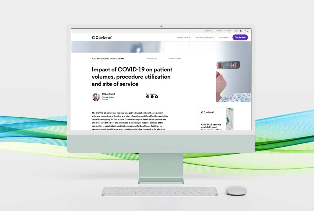
Although the COVID-19 pandemic negatively impacted the healthcare industry during lockdowns, we also saw many markets recover quite rapidly within 2020. This report just published outlines the impact COVID-19 pandemic had on healthcare patient volumes, procedure utilization and sites of service, and how the effect varied by procedure urgency.
Did you miss the webinar on Fluorescence Guided Surgery? Log in for free to hear from the experts
Listen back to the great webinar on FGS last week and what one surgeon described as “Essential
technology to reduce complications, improve the outcomes we get in foregut and bariatric surgery”.
Click here to log in: https://aischannel.com/live-surgery/fluorescence-imaging/
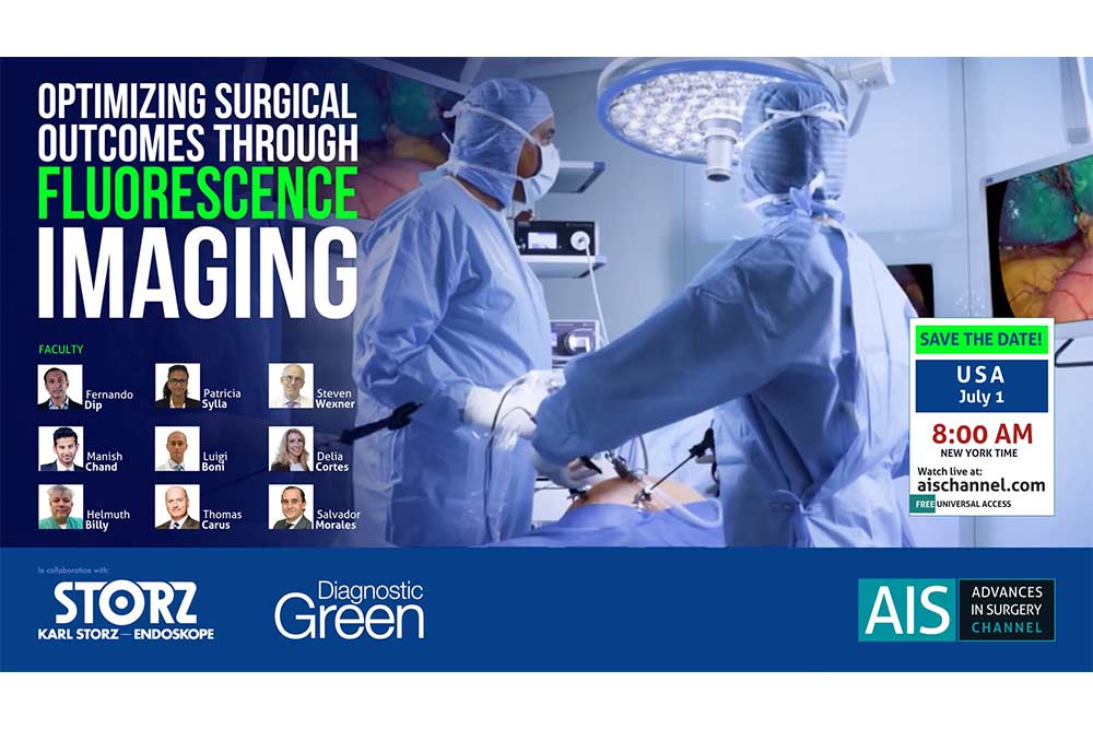
Want to learn from surgeons who are expert in Fluorescence Guided Surgery? Then log into a free webinar this Thursday July 1
Meta-analysis of indocyanine green fluorescence imaging-guided laparoscopic hepatectomy
This meta-analysis was conducted to systematically evaluate the short-term efficacy and safety of indocyanine green (ICG) fluorescence imaging-guided laparoscopic hepatectomy.
This meta-analysis included 6 studies comprising 417 patients with liver disease. The meta-results showed that compared to the control group, ICG fluorescence imaging-guided laparoscopic hepatectomy can significantly shorten the operative time [weighted mean differences (WMD) = -20.81, 95% CI, -28.02–13.59, p = 0.000], reduce intraoperative bleeding [WMD = -108.16, 95% CI, -127.88–88.44, p = 0.000], shorten hospital stay [WMD= -1.23,95% CI, -1.50–0.95, p = 0.000], and reduce the incidence of postoperative complications [OR = 0.49,95% CI, 0.26-0.91, p = 0.025]. There were no differences in blood transfusion, hilar occlusion time, and surgical margin.
Conclusion: The application of ICG fluorescence imaging technology in laparoscopic hepatectomy can effectively reduce the operative time, blood loss, hospital stay and the incidence of postoperative complications.
Advances in Surgery Live Webinar Event focusing on Fluorescence Imaging – SAVE THE DATE – JULY 1
We are very excited to bring you this webinar event focusing on fluorescence imaging on July 1. The webinar will bring together a group of eminent surgeons from around the world, expert in fluorescence guided surgery.
Check out https://aischannel.com/live-surgery/fluorescence-imaging/ for more information.
Coronavirus tracker: More than 96% of physicians fully vaccinated against COVID-19, AMA survey shows
The national AMA survey polled 300 physicians, including primary care doctors and specialists, between June 3-8. It’s the first survey to specifically collect data on practicing physicians’ COVID-19 vaccination rates, according to the AMA. The survey results show an increase of more than 20% for physicians who have been fully vaccinated for COVID-19 compared to a May 2021 Medscape poll. “Practicing physicians across the country are leading by example, with an amazing uptake of the COVID-19 vaccines,” said AMA President Susan R. Bailey, M.D. in a statement. There were no significant differences in physician vaccination rates across various demographic groups, including: PCP vs Specialist, region, gender, age, and race. However non-Hispanic physicians (97%) were more likely to have been vaccinated than Hispanic physicians (84%).
https://www.ama-assn.org/system/files/2021-06/physician-vaccination-study-topline-report.pdf
COVID-19: Pandemic Surgery Guidance – basis for handling surgeries in future potential crises
Various scientists and clinicians from disparate specialties provided a Pandemic Surgery. These included recommendations regarding surgical response from Society of American Gastrointestinal and Endoscopic Surgeons (SAGES) and the European Association for Endoscopic Surgery (EAES). Guidance for surgical procedures by distinct surgical disciplines such as numerous cancer surgery disciplines, cardiothoracic surgery, ENT, eye, dermatology, emergency, endocrine surgery, general surgery, gynecology, neurosurgery, orthopedics, pediatric surgery, reconstructive and plastic surgery, surgical critical care, transplantation surgery, trauma surgery and urology, performing different surgeries, as well as laparoscopy, thoracoscopy and endoscopy. The present Pandemic Surgery Guidance could even serve as the basis for other future potential pathogen crises yet to come.
https://www.researchgate.net/publication/340548953_COVID-19_Pandemic_Surgery_Guidance
Optical Coherence Tomography Angiography Compared with Multimodal Imaging for Diagnosing Neovascular Central Serous Chorioretinopathy
A prospective cross-sectional study to assess the diagnostic accuracy of optical coherence tomography angiography (OCTA) compared with multimodal imaging for CNV in CSC eyes and to determine the features that predicted CNV.
Methods: Consecutive CSC patients were recruited from retina clinic. The reference standard for CNV was determined by interpretation of multimodal imaging with OCTA, structural OCT line scan, fluorescein angiography (FA), indocyanine green angiography (ICGA), ultra-widefield fundus photography and fundus autofluorescence (FAF). Two independent masked graders examined OCTA without FA and ICGA to diagnose CNV. Univariate and multivariate analyses were performed to evaluate factors associated with CNV.
Conclusions: There is discordance between OCTA and multimodal imaging in diagnosing CNV in CSC. This study demonstrated the caveats in OCTA interpretation, such as small extrafoveal lesions and retinal pigment epithelial alterations. Comprehensive interpretation of OCTA with dye angiography and structural OCT is recommended.
The role of indocyanine green cholangiography in minimally invasive surgery, published June
This paper examines Near-infrared fluorescent cholangiography (NIFC) using indocyanine green (ICG) to aid in the identification of extrahepatic biliary anatomy.
Evidence synthesis: Several factors can influence the quality of the fluorescence imaging, including the dose and timing of ICG injection, liver function, the thickness of fatty tissue and the presence of inflamed tissues due to acute pathology. Various devices tested also have a different sensitivity to the fluorescence signal. RCTs showed fluorescence cholangiography were comparable to traditional intraoperative cholangiogram in visualizing the extrahepatic biliary anatomy.
Conclusions: NIFC is demonstrated as a safe, non-irradiating technique to identify and aid in the visualization of extrahepatic biliary anatomy. Laparoscopic cholecystectomy with real-time NIFC enables a better visualization and identification of biliary anatomy and therefore it is potentially as a means of increasing the safety of laparoscopic cholecystectomy. Whether this translates into reducing complication rates must still be determined. The dosage and timing of the intravenous administration of ICG relative to the operative procedure still requires optimization to ensure reliable images.
Recent case report on “Use of Indocyanine Green Fluorescent Imaging in the Assessment of a Tongue Flap After Lateral Hemiglossectomy”
Indocyanine green (ICG) angiography is a real-time imaging modality that can be used to assess intraoperative tissue perfusion. ICG dye has proven to be feasible, safe, and cost-effective, especially for muscle flaps during complex reconstructions. To our knowledge, we discuss the first use of ICG angiography for the real-time assessment of a tongue flap following left lateral hemiglossectomy. ICG angiography showed excellent perfusion of the tongue and tongue flap, which subsequently led to an uncomplicated postoperative recovery.
Conclusion: The use of ICG angiography for the real-time assessment of blood flow is a valuable and feasible tool for the intraoperative assessment of perfusion in extended hemiglossectomies with local flap reconstruction. It may be especially beneficial in cases involving complex reconstructions, and when regional organ hypoperfusion is suspected.

