Use of Indocyanine Green for Injection USP (ICG) in
Ophthalmology
Indocyanine Green Angiography (ICGA) is the gold standard in diagnosing a number of serious eye conditions and is a
key diagnostic tool used by ophthalmology specialists worldwide.
ICGA is particularly useful in the differential diagnosis of Polypoidal Choroidal Vasculopathy (PCV), Central Serous Chorioretinopathy (CSCR), and Retinal Angiomatous Proliferation (RAP), which can be misdiagnosed as nAMD (Neovascular Age-related Macular Degeneration)1.

Key Features
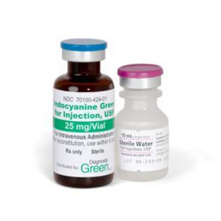
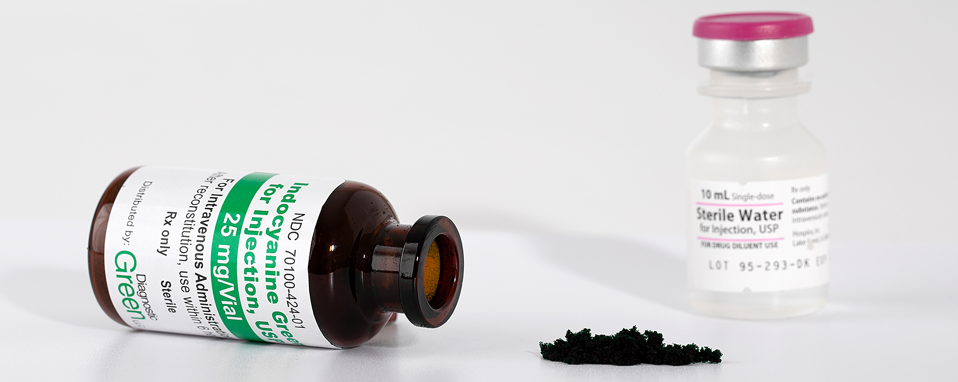
Indocyanine Green for Injection USP (ICG)) is a tricarbocyanine dye with both hydrophilic and lipophilic properties.
The retention of ICG in the fenestrated choroidal circulation, combined with its low permeability, makes ICG angiography ideal for viewing the choroidal blood vessels.
Once injected, the ICG binds to plasma proteins and quickly circulates to the choroid layer, delineating the choroidal veins within 15-20 secs.
Indocyanine Green for Injection USP is cleared exclusively through the liver and then excreted through the bile. It does not undergo metabolism. Indocyanine Green for Injection USP has an excellent safety profile and adverse reactions occur very rarely (<1/10,000).
Using ICGA at initial presentation helps identify disorders of the choroidal circulation, allowing differential treatment approaches that may improve outcomes and safety for patients.
- Investigation of complex posterior uveitis and white dot syndromes
- Assessment of patients with “wet” AMD where the presence of polypoidal choroidal vasculopathy (PCV) is in question
- The assessment of choroidal hyperpermeability in patients with central serous chorioretinopathy
Check out the Product Information Leaflet (ICG) here
| ICGA | OCTA |
|---|---|
| ICGA fluorescence can penetrate blood, fluid and retinal pigment epithelium to reveal underlying abnormalities of the inner choroidal vasculature and is essential for making a definitive diagnosis of PCV. | Extremely motion sensitive, requiring a patient to fixate on precise point for several seconds. Patient compliance required, which is often difficult, particularly for older patients. |
| Excellent visualisation within minutes, of the medium & large choroidal vessels. | OCTA takes more time than structural scans and requires trade-offs in flow resolution, scan quality and speed. |
| ICGA is beneficial in the differential diagnosis of PCV, Chronic CSC, and RAP. | Limited field of view leading to a greater likelihood that lesions may be missed. |
| ICGA has been shown to optimise detection of capillary macro aneurysms in longstanding diabetic macular edema (DME) or retinal vein occlusion (RVO). | Failure to recognise OCTA Projection Artifact (blood vessels seem at erroneous location), may lead to inaccurate clinical assessment. |
| A recent study demonstrated that late leakage in ICGA occurred in all RAP cases*. | Image processing for OCTA can alter blood vessel appearance through egmentation defects, and image display software can lead to false impressions of vessel location and density |
| Duration of ICGA procedure only 15 20mins, very quick analysis. | The analysis of these images is time-consuming – may involve many hours of post hoc manual segmentation work, which may be difficult to accommodate during daily medical work routines. |
*Ravera V, et al.. RETINAL ANGIOMATOUS PROLIFERATION DIAGNOSIS: A Multiimaging Approach. Retina. 2016;36(12):2274– 2281.