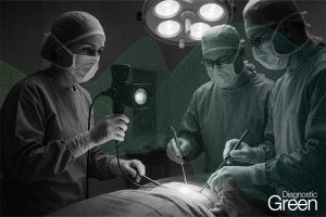
BACKGROUND: The aim of this study was to assess the effect of indocyanine green (ICG) fluorescence imaging combined with laparoscopic ultrasound in laparoscopic microwave ablation of liver cancer.
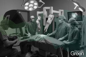
MATERIAL AND METHODS: This study retrospectively analyzed 61 patients who underwent laparoscopic microwave ablation of liver cancer, including laparoscopic microwave ablation with and without ICG fluoroscopy.
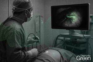
RESULTS: The operative times, ablation times, postoperative hospital stay, postoperative complication rate, hospitalization cost, postoperative liver function changes, and postoperative overall survival were similar between the 2 groups, but there was a statistically significant difference in recurrence-free survival (P<0.05). A total of 5 lesions were found in the fluorescence laparoscopy group that were not found by preoperative imaging, while no new lesions were found in the ordinary laparoscopy group. Fluorescence laparoscopy has obvious advantages over ordinary laparoscopy in finding small lesions that were not found before surgery. In terms of complete ablation rate, 3 patients in the ordinary laparoscopy group and 1 patient in the fluorescence laparoscopy group were judged to be incompletely ablated and were ablated again at 1 month after the operation.
CONCLUSIONS: For small hepatocellular carcinoma with severe liver cirrhosis and located on the liver surface, fluorescence laparoscopy can better reveal the location and boundary of the tumor, and fluorescence laparoscopy can detect tiny lesions that cannot be detected by preoperative imaging. The combination of fluorescence laparoscopy and microwave ablation has a good effect on the treatment of small hepatocellular carcinoma located on the surface of the liver that is difficult to distinguish.