In cases of locally advanced oral tongue cancer, a wide surgical excision followed by appropriate reconstruction, preferably using free flaps, is recommended.The intricate nature of the lingual anatomy cannot be “replaced in kind;” thus, there is the need for optimization of tissue preservation when resecting tumors. Although the tongue receives ample vascular supply from the lingual artery and its branches, including the sublingual artery, the dorsal artery of the tongue, and the deep lingual artery, there are also minor contributions to its blood supply from adjacent vascular territories. These include the anastomosis between the ascending palatal artery and the dorsal tongue artery, as well as the connection between the submental artery and the deep tongue artery and sublingual artery.
Additionally, there are anastomotic networks present in the tongue base and dorsal surface. However, the avascular midline septum is only occasionally crossed by submucosal branches (the longitudinal median orientation of the superficial vasculature), and the specific vascular territories of each arterial branch may lead to a compromised vascular supply at the edges of the remaining tongue after tumor resection. Furthermore, in head and neck reconstructions, complications such as dehiscence and fistula are observed in 27.2% and 6.9% of recipient sites, respectively. To mitigate these complications, significant attention has been given to the vascularization of flaps, but the quality of the recipient site itself has received less emphasis. Partial tongue necrosis can occur, leading to wound dehiscence. In head and neck surgery, indocyanine green (ICG) angiography is a commonly used technique that has demonstrated its usefulness in assessing tissue perfusion, aiding in surgical decision-making, minimizing complication rates in microvascular free, pedicled, and random flaps, and predicting fistula occurrence following salvage surgery.
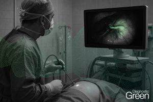
The objective of this video article was to provide a detailed description of the steps involved in utilizing ICG angiography for assessing recipient site perfusion after glossectomy with the primary goal of minimizing wound healing complications, especially when performing free flap reconstruction. (See Video 1 [online], which displays case presentation and set up) (See Video 2 [online], which displays indocyanine green injection and perfusion assessment of the remaining tongue) (See Video 3 [online], which displays remaining tongue resection and edge bleeding control perfusion) (See Video 4 [online], which displays assessment of the flap and functional outcomes)
ICG angiography for assessing the viability of the remaining tongue after glossectomy is simple to implement and takes only a few extra minutes to perform. This approach has the potential to reduce postoperative wound healing complications in head and neck reconstruction.