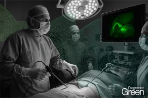
Near-infrared (NIR) fluorescence imaging with indocyanine green (ICG) has gained popularity in pediatric surgery as it has in general surgery. In addition, a water-jet dissector (WJD) has been successfully introduced in adult hepatic surgery. Tissue structures are dissected selectively and gently by the WJD. However, there have been no reports of hepatic resection for pediatric patients using a WJD. We applied NIR fluorescence imaging with ICG to visualize the resection line of the liver and used a WJD for liver parenchyma dissection in pediatric hepatoblastoma.
The patient was a 3-year-old girl with a large liver tumor. Enhanced computed tomography revealed a liver tumor (maximum diameter: 120 mm) in the right lobe and three small lung metastases. The liver tumor was diagnosed as hepatoblastoma (PRETEXT 2) based on an open biopsy. We performed right hepatectomy after neoadjuvant chemotherapy. The right lobe was mobilized from the diaphragm, and then intraoperative ultrasound was performed to detect the localization of the tumor and its proximity to the vascular structures. We detected the right hepatic artery (RHA), right portal vein (RPV), and right hepatic vein (RHV).
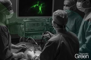
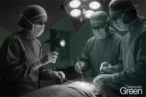
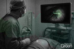
The middle hepatic vein was not involved. After ligation of the RHA and RPV to selectively control the right lobe inflow, ICG was administered intravenously and observed by an NIR endoscope. The resection line was clearly visualized by overlaying images in comparison to conventional demarcation line detection. Then, we used a WJD to dissect the parenchyma. Small vessels were divided from parenchymal tissue and were clearly visible. We resected them after clamping with metal clips. Finally, the RHV was transected by a linear stapler, and right hepatectomy was completed with 25 ml of blood loss. There was no postoperative hemorrhage. We performed hepaticojejunostomy because of stricture of the common bile duct on postoperative day 302.
The patient was discharged after adjuvant chemotherapy. NIR imaging clearly showed the resection line. The WJD automatically separated, and thus made visible, the more resistant duct and vessel structures from the parenchyma. The combined use of NIR imaging and WJD was useful for pediatric hepatectomy.