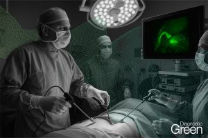
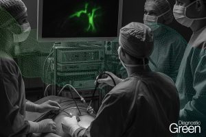
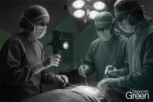
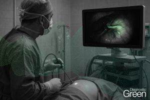
Purpose: To evaluate the safety and efficacy of indocyanine green fluorescence imaging for real-time guidance of laparoscopic thermal ablation in patients with liver cancer.
Materials and methods: A total of 27 patients with 40 liver lesions underwent fluorescence-assisted laparoscopic ablation between January 2020 to March 2023. The sensitivity of indocyanine green (ICG)-fluorescence imaging, technique effectiveness rate and complications of fluorescence-assisted laparoscopic thermal ablation were evaluated.
Results: In total, 33 out of the 40 lesions were identified by ICG-fluorescence imaging technique, with the sensitivity of 82.5%. The sensitivity of ICG-fluorescence imaging of tumor detection in liver surface of parenchyma was significantly higher than that in the deeply located hepatic parenchyma (96.8% vs 33.3%, p = 0.002). ICG-fluorescence imaging procedures detected 4 lesions that cannot be seen on intraoperative ultrasound. It provides clear demarcation lines on the hepatic surface. Technical success is achieved if the necrotic zone had at least a 5 mm ablative margin around the outer edge of the ICG-fluorescence image. Technical success of fluorescence laparoscopic radiofrequency ablation (FLRFA) and fluorescence laparoscopic microwave ablation (FLMWA) was 100% (27/27). Technical effectiveness is defined by the complete necrotic lesions of the local tumor tissue during follow-up. According to the CT/MRI one month after FLRFA or FLMWA, the technical efficacy rate was 92.5% (37/40) and local tumor progression occurred in 7.5% (3/40) of the enrolled lesions. During the follow-up period, no major complications were observed.
Conclusion: ICG-fluorescence imaging guided laparoscopic thermal ablation was feasible, safe and effective.