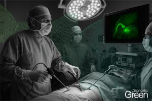
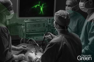
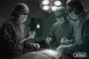
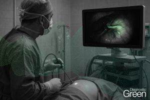
Background: Ultrasonography (US) is widely used for pre-operative detection of liver tumors. However, US does not have high resolution and very small tumors, tumors located near the liver surface, or those in cirrhotic livers are often not detected. Case Report: A 47-year-old woman with a previous surgery for sigmoid colon cancer (T3N1bM0 Stage3b) showed a liver tumor on the surface of segment 2 by contrast-enhanced computed tomography (CT) and gadoliniumethoxybenzyldiethlenetriaminepen-taacetic acid (Gd-EOB-DTPA) magnetic resonance imaging (MRI). Laparoscopic liver resection was performed under fluorescence guidance (using Indocyanine Green, ICG). Pathological examination showed a pseudotumor with negative margins. The present study showed that ICG can allow the detection of tumors in the liver that are invisible with IOUS. Furthermore ICG fluorescence has a higher detection rate for small tumors or tumors near the surface of the liver than IOUS.
Conclusion: ICG fluorescence imaging can allow visualization of liver tumors that are undetectable on US.