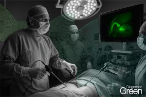
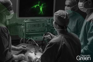
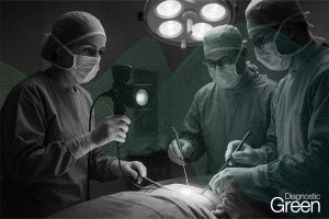
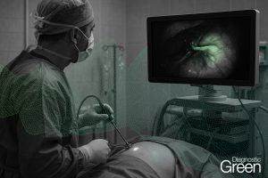
Revision surgery for the complications after repair of esophageal atresia is often complex because of previous surgeries and chest infections and thus requires surgical expertise. This study describes surgical experiences with the use of indocyanine green (ICG) fluorescence imaging localization-assisted thoracoscopy during revision surgery, including recurrent tracheoesophageal fistula (rTEF) (8 cases, one of which was esophageal-pulmonary fistula) and delayed esophageal closure (1 case).
We performed fistula repair and esophageal reconstruction according to the indications of ICG. The application of this method avoids the excessive trauma caused by freeing the trachea and esophagus. Contrast imaging taken one week and one month after surgery indicated no spillover of the contrast agent from the esophagus, except in 1 case. Indocyanine green fluorescence imaging localization-assisted thoracoscopy is worth promoting for revision surgery after esophageal atresia repair.
In summary, the use of indocyanine green fluorescence localization-assisted thoracoscopy for revision surgery to treat complications after esophageal atresia repair is a safe and reliable method that deserves promotion.
https://bmcgastroenterol.biomedcentral.com/articles/10.1186/s12876-022-02444-1