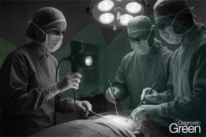
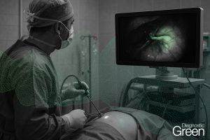
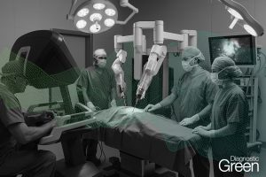
The wide availability of near infrared light sources in interventional medical imaging stacks enables non-invasive quantification of perfusion by using fluorescent dyes, typically Indocyanine Green (ICG). Due to their often leaky and chaotic vasculatures, intravenously administered ICG perfuses through cancerous tissues differently. We investigate here how a few characteristic values derived from the time series of fluorescence can be used in simple machine learning algorithms to distinguish benign lesions from cancers. These features capture the initial uptake of ICG in the colon, its peak fluorescence, and its early wash-out. By using simple, explainable algorithms we demonstrate, in clinical cases, that sensitivity (specificity) rates of over 95% (95%) for cancer classification can be achieved.