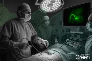
Anastomotic leakage (AL) remains a prevalent and life-threatening complication after esophagectomy. Gastric tube perfusion assessment using indocyanine green fluorescence imaging (ICG-FI) has been published in several studies and appears to be a promising tool to reduce AL rates by changing the surgical approach, namely by an intraoperative evaluation of the anastomosis localization.
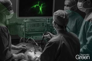
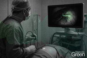
In this study, gastric tube perfusion was quantified by using ICG-FI in 20 high-risk patients undergoing esophagectomy. From a time-dependent fluorescence intensity curve, the following three parameters were evaluated: slope of fluorescence intensity (SFI), background subtracted peak fluorescence intensity (BSFI), and time to slope (TTS). The values between pyloric region and tip showed a similar downward trend and SFI and BSFI significantly correlated with the distance to the pyloric region. SFI and BSFI were significantly decreased at the tip of the gastric tube. The placement of anastomosis in an area with homogenous fluorescence pattern was correlated with no AL in 92.9% of cases. An inhomogeneous fluorescence pattern at anastomotic site was a risk factor for the occurrence of an AL (p < 0.05). Reduction of perfusion up to 32% using SFI and up to 23% using BSFI was not associated with AL.
Conclusion: ICG-FI can be used to quantify the gastric tube perfusion by calculating SFI, BSFI, and TTS. The anastomosis should be created in areas with homogeneous fluorescence pattern. A reduction in blood flow of up to 32% can be accepted without causing an increased rate of insufficiency.