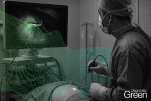
Background: Indocyanine-Green Fluorescence Angiography (ICG-FA) is widely used in reconstructive surgery, providing real-time visualization of flap perfusion. Accurate assessment of perfusion is especially critical in lower extremity reconstructions, where complications like necrosis and venous congestion can lead to poor outcomes, including amputation. Although ICG-FA is commonly available, its interpretation remains subjective and heavily reliant on the surgeon’s experience. These challenges underline the importance of integrating objective, data-driven assessment tools into surgical practice.
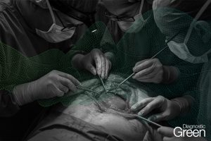
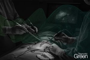
Methods: As part of a larger, ongoing prospective study, three illustrative cases of lower extremity reconstructions using perforator-based fasciocutaneous flaps were selected. Intraoperative ICG-FA was performed using a surgical microscope with integrated fluorescence imaging. Fluorescence-time-curves (FTCs) were generated using specialized software, and associated quantitative perfusion parameters were compared across three cases: two patients with perfusion-related complications and one patient without complications.
Results: Intraoperative clinical assessment appeared satisfactory in all cases, and no changes in surgical management were made based on the subjective interpretation of ICG-FA. In contrast, quantitative analysis of ICG-FA revealed abnormal perfusion patterns in the two flaps that developed complications, identifying perfusion deficits not evident through conventional assessment.
Conclusion: These findings suggest that FTCs derived from ICG-FA data can predict perfusion-related complications. Integrating quantitative ICG-FA analysis into clinical practice may yield a significant advancement in reconstructive surgery, especially in lower extremity reconstructions. Clinical trial name ICG Indocyanine Green in Reconstructive Surgery (ICG-R).ClinicalTrials.gov IDNCT06129669 (https://clinicaltrials.gov/study/NCT06129669?cond=NCT06129669&rank=1 ).