Introduction: Paraduodenal hernias are rare congenital anomalies that can lead to acute bowel obstruction and strangulation. Laparoscopic management of these complex cases in emergency settings remains challenging, particularly when bowel ischemia is present.
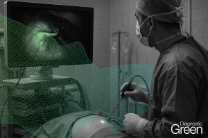
Case presentation: We report a case of a 56-year-old woman presenting with acute small bowel obstruction due to a strangulated left paraduodenal hernia. Emergency laparoscopic surgery revealed ischemic bowel segments within the hernia sac. We utilized indocyanine green (ICG) fluorescence imaging to assess bowel perfusion intraoperatively, guiding our decision for bowel resection. The procedure involved hernia reduction, resection of non-viable bowel, and primary anastomosis, followed by hernia defect closure. Despite encountering a small bowel injury during reduction, we successfully completed the procedure laparoscopically.
Discussion: This case demonstrates the feasibility of laparoscopic management for complicated paraduodenal hernias with bowel strangulation in emergency settings. The use of ICG imaging for real-time perfusion assessment represented a novel application in this context, aiding in the precise identification of ischemic bowel segments requiring resection.
Conclusion: Laparoscopic repair of strangulated paraduodenal hernias is feasible and effective, even in emergency scenarios. The integration of advanced imaging techniques like ICG fluorescence may enhance intraoperative decision-making, particularly in assessing bowel viability. This approach potentially reduces the extent of bowel resection and improves outcomes in these complex cases.
https://www.sciencedirect.com/science/article/pii/S2210261224013476?via%3Dihub


