This website contains information intended exclusively for physicians, pharmacists, and other qualified healthcare professionals.
Please confirm your status to continue
By clicking “Yes,” you confirm that you are a licensed healthcare professional and acknowledge that this content is intended for professional use only and not intended for use by the general public.
IC-Flow™ Imaging System is a compact and easy to use handheld device that visualizes and records tissue fluorescence.
Point of Care System
Compact and easily portable
Easy to use
Data transfer to USB stick
Fluorescence image on integrated display
No additional equipment required
Operating Room System
Portable cart system
Integrated system with monitor
External USB for official recording of patient data
Display of fluorescence image on both touchscreen and monitor
Unique Benefits
Backed by Diagnostic Green, the global supplier of ICG and 30 years’ experience in perfusion imaging, the IC-Flow™ Imaging System, is a small, yet powerful imaging device for you and your surgical team.
Fast boot-up time
Simple and easy to use with minimal training required
Flexible configurations to meet speciality needs
Full OR System
Portable Point of Care System
Safe use with the benefit of LED rather than laser light source
Minimal maintenance
Minimal service requirements
Testimonials
“The cost benefit of the IC-Flow™ Imaging System
is more than justified. This portable camera is easy to
use and provides an efficient fluorescence image.”
IC Flow Imaging system is “user friendly, affordable and compact”
The IC-Flow Imaging system “generates images of fluorescence in order to make a conclusion and a treatment decision”
Better Outcomes, Reduced Costs
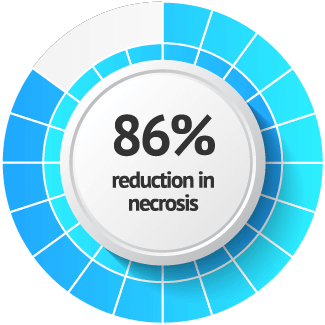
Breast Reconstruction Surgery1
(Clinical assessment 26.5% vs laser-assisted ICG aniography 5%)
Published literature has well established the significant costs associated with surgical complications including those related to post-mastectomy breast reconstruction and mastectomy flap and free flap survival1,2,3. In a USA study, the average excess cost associated with a single case of flap necrosis was calculated to be almost $100K per patient3. Many studies have shown that the use of ICG in conjunction with a visualization system, like IC-FlowTM Imaging System, results in reduced failure rates, reduced healthcare costs, and overall better outcomes for patients4,5,6.
Chatterjee et al. Plast Reconstr Surg. 2013;131(5):693e–701e.
Harless et al. Breast J. 2016;22(3):274–81.
An Outcome Analysis of Intraoperative Angiography for Postmastectomy Breast Reconstruction. Duggal CS. Aesthetic Surgery Journal. 2014; 34(1):61-5
Indocyanine green fluorescence angiography for free flap monitoring: A pilot study, J Craniomaxillofac Surg. 2016 Nov;44(11):Hitier M, Cracowski JL, Hamou C, Righini C, Bettega G
ICG angiography in immediate and delayed autologous breast reconstructions: preoperative evaluation and postoperative outcomes, J Plast Surg Hand Surg. 2018 Oct;52(5); Alstrup T, Christensen BO, Damsgaard TE.
Use of Intraoperative Fluorescent Angiography to Assess and Optimize Free Tissue Transfer in Head and Neck Reconstruction; Journal of Maxillofacial surgery, August 2013; Volume 71, Issue 8, p1305-1480; J. Marshall Green, Shane Thomas, DO, Jennifer Sabino, MD, Robert Howard, MD, Patrick Basile, MD⋮, Steven Dryden, DDS, Chris Crecelius, DDS, Ian Valerio, MD, MS, MBA.
The information on this page is only intended for residents of the chosen country. Please confirm that you are declaring and confirming that you are a healthcare professional and have read and understood this disclaimer.