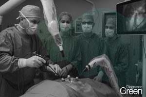
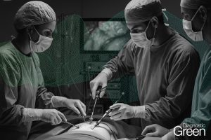
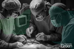
Pure laparoscopic donor hepatectomy (PLDH) has become a standard practice for living donor liver transplantation in expert centers. Accurate understanding of biliary structures is crucial during PLDH to minimize the risk of complications. This study aims to develop a deep learning-based segmentation model for real-time identification of biliary structures, assisting surgeons in determining the optimal transection site during PLDH.
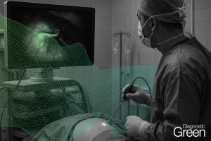
A single-institution retrospective feasibility analysis was conducted on 30 intraoperative videos of PLDH. All videos were selected for their use of the indocyanine green near-infrared fluorescence technique to identify biliary structure. From the analysis, 10 representative frames were extracted from each video specifically during the bile duct division phase, resulting in 300 frames. These frames underwent pixel-wise annotation to identify biliary structures and the transection site.
A segmentation task was then performed using a DeepLabV3+ algorithm, equipped with a ResNet50 encoder, focusing on the bile duct (BD) and anterior wall (AW) for transection. The model’s performance was evaluated using the dice similarity coefficient (DSC). The model predicted biliary structures with a mean DSC of 0.728 ± 0.01 for BD and 0.429 ± 0.06 for AW. Inference was performed at a speed of 15.3 frames per second, demonstrating the feasibility of real-time recognition of anatomical structures during surgery. The deep learning-based semantic segmentation model exhibited promising performance in identifying biliary structures during PLDH. Future studies should focus on validating the clinical utility and generalizability of the model and comparing its efficacy with current gold standard practices to better evaluate its potential clinical applications.