Indocyanine Green Labeling of Tumors in the Liver Recurring After Radiofrequency Ablation Enables Complete Resection by Fluorescence-guided Surgery

Background/aim: Radiofrequency ablation (RFA) is used to treat primary and metastatic tumors in the liver. However, local recurrence after RFA is frequent and subsequent salvage hepatectomy is often ineffective due to difficulty in visualization of tumor margins. Patients and methods: In the present retrospective clinical trial, seven patients from the Department of General and Gastro-enterological Surgery, Showa University School of Medicine underwent salvage hepatectomy for recurrent hepatocellular carcinoma (HCC) (n=2), colorectal liver metastasis (n=4) and lung-carcinoid liver metastasis (n=1), after RFA, between 2011 and 2020. Tumors were labeled with indocyanine green (ICG) and resected under fluorescence guidance. Resected specimens were evaluated under fluorescence microscopy as well as by standard histopathological techniques.

Results: Pathological findings revealed negative tumor margins in all patients after fluorescence-guided surgery. Six of seven resected tumors had a fluorescent rim, including both HCC and liver metastasis. Fluorescence microscopy demonstrated that viable cancer tumor cells were located only on the inside of the fluorescent rim, and no malignant cells were detected within the fluorescent rim surrounding the tumor. Fluorescence microscopy showed that the tumor margin was secured if the fluorescence signal was completely resected.

Conclusion: The present results demonstrate that ICG labeling of liver tumors recurring after RFA enabled complete resection under fluorescence guidance.

https://pubmed.ncbi.nlm.nih.gov/35220226/

Efficacy of Near-Infrared Fluorescence-Guided Hepatectomy for the Detection of Colorectal Liver Metastases: A Randomized Controlled Trial

Background: The application of indocyanine green fluorescence-guided hepatectomy for liver metastases from colorectal cancer is in the preliminary stage of clinical practice; thus, its efficacy needs to be determined. This study compared the number of intrahepatic colorectal liver metastases detected intraoperatively and postoperative recovery data between patients who underwent traditional hepatectomy (nonindocyanine green group) and traditional hepatectomy plus intraoperative indocyanine green fluorescence imaging (indocyanine green group).

Overall, we recruited 80 patients, among whom 72 eligible patients were randomly assigned. After allocation, 64 patients, comprising 32 in each group, underwent the allocated intervention and follow-up. Compared with the nonindocyanine green group, the mean number of intrahepatic colorectal liver metastases identified intraoperatively in the indocyanine green group was significantly greater (mean [standard deviation], 3.03 [1.58] vs 2.28 [1.35]; p = 0.045), the postoperative hospital stay was shorter (p = 0.012) and the 1-year recurrence rate was also lower (p = 0.017). Postoperative complications and 90-day mortality were comparable, with no statistical differences.

Conclusions: Indocyanine green fluorescence imaging significantly increases the number of intrahepatic colorectal liver metastases identified and reduces postoperative hospital stay and 1-year recurrence rate without increasing hepatectomy-related complications and mortality rates.

https://pubmed.ncbi.nlm.nih.gov/35213433/

Diagnosis and Treatment of Primary Inflammatory Choriocapillaropathies (PICCPs): A Comprehensive Overview

Purpose: Primary inflammatory choriocapillaropathies (PICCPs) belong to a group of intraocular inflammatory diseases with the common characteristic of inflammatory choriocapillaris hypo- or non-perfusion as the main clinicopathological mechanism. The purpose of our article is to describe clinical characteristics and multimodal imaging, that can help the diagnosis and treatment of PICCPs.

Diagnosis is mostly based on multimodal imaging and especially on indocyanine green angiography (ICGA), fundus autofluorescence (FAF) and spectral-domain optical coherence tomography (SD-OCT)/OCT-angiography (OCT-A). ICGA shows the typical pattern of patchy lobular hypofluorescence reflecting hypo- or non-perfusion of the choriocapillaris that can also take the aspect of geographic areas in the more severe forms. Treatment depends on the severity of the disease and goes from observation in MEWDS and some mild cases of APMPPE, to oral corticosteroid and/or immunomodulator agents in the more severe conditions of APMPPE and MFC and SC cases. Close multimodal monitoring is crucial in order to introduce or adjust treatment.

Conclusion: PICCPs are resulting from one common clinicopathological mechanism, inflammatory choriocapillaris hypo- or non-perfusion. ICGA findings are essential for the diagnosis and follow-up of PICCPs, but non-invasive methods such as FAF and SD-OCT/OCT-A also have their role especially in follow-up of the diseases. Treatment should be individualized according to the pathology and the evolution of lesions.

https://pubmed.ncbi.nlm.nih.gov/35208488/

Hospital revenues, delayed elective surgeries top trends to watch over next year: survey

A poll of more than 2,300 healthcare leaders by Definitive Healthcare found more than 36% of respondents called the loss of revenue in hospitals the single biggest trend to watch in the industry in the year ahead.  Nearly a quarter of those polled said the implications of delayed care including surgeries would be the most crucial area of change because of the potential ripple effects across the industry if patients’ deferred preventive and critical care resulted in serious health implications, they said.

“The concern, of course, is that as we get into the rest of the year, there is going to be a pent-up demand for those surgeries,” Todd Bellemare, Definitive Healthcare’s vice president of professional services, told Fierce Healthcare. “There will be a bit of a bounce back when people start going back to the hospital to schedule these surgeries.”

https://www.definitivehc.com/resources/healthcare-insights/predicting-covid-19-long-term-effects

Multisensor perfusion assessment cohort study: Preliminary evidence toward a standardized assessment of indocyanine green fluorescence in colorectal surgery

Traditional methods of assessing colonic perfusion are based on the surgeon’s visual inspection of tissue. Fluorescence angiography provides qualitative information, but there remains disagreement on how the observed signal should be interpreted. It is unclear whether fluorescence correlates with physiological properties of the tissue, such as tissue oxygen saturation.

The aim of this study was to correlate fluorescence intensity and colonic tissue oxygen saturation. In total, 18 patients were included. Maximal fluorescence intensity occurred at a mean of 101 seconds after indocyanine green injection. The correlation coefficient was 0.73 (95% confidence interval of 0.65–0.79) with P < .0001, showing a statistically significant strong positive correlation between normalized fluorescence intensity and tissue oxygen saturation. The use of time averaging improved the correlation coefficient to 0.78.

Fluorescence intensity is a potential surrogate for tissue oxygenation. This is expected to lead to improved decision making when transecting the bowel and, consequently, a reduction in anastomotic leak rates. A larger, phase II study is needed to confirm this result and form the basis of computational algorithms to infer biological or physiological information from the fluorescence imaging data.

https://www.surgjournal.com/article/S0039-6060(21)01244-7/fulltext

Role of Indocyanine Green Fluorescence Imaging in Minimally Invasive Resection of Colorectal Liver Metastases

Indocyanine green (ICG) fluorescence imaging is an easy and reproducible method to detect hepatic lesions, both primary and metastatic. This review reports the potential benefits of this technique as a tactile mimicking visual tool and a navigator guide in minimally invasive liver resection of colorectal liver metastases (CRLM). PubMed and MEDLINE databases were searched for studies reporting the use of intravenous injection of ICG before minimally invasive surgery for CLRM.

The search was performed for publications reported from the first study in 2014 to April 2021. The final review included 13 articles: 6 prospective cohort studies, 1 retrospective cohort study, 3 case series, 1 case report, 1 case-matched study, and 1 clinical trial registry. The administered dose ranged between 0.3 and 0.5 mg/kg, while timing ranged between 1 and 14 days before surgery. CRLM detection rate ranged between 30.3% and 100% with preoperative imaging (abdominal computed tomography/magnetic resonance imaging), between 93.3 and 100% with laparoscopic ultrasound, between 57.6% and 100% with ICG fluorescence, and was 100% with combined modalities (ICG and laparoscopic ultrasound) with weighted averages of 77.42%, 95.97%, 79.03%, and 100%, respectively.

ICG fusion imaging also allowed to detect occult small-sized lesions, not diagnosed preoperatively. In addition, ICG is effective in real-time assessment of surgical margins by evaluating the integrity of the fluorescent rim around the CRLM.

https://pubmed.ncbi.nlm.nih.gov/35180735/
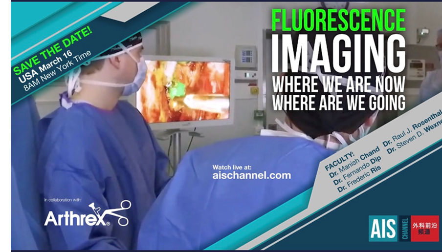
Reduce Pre-Surgery Anxiety by Getting to Know Your Options – Sponsored by the ISFGS

“FGS is one of the most revolutionary technologies in the field of surgery today,” says Dr. Raul Rosenthal, regional chairman for the Digestive Disease and Surgical Institute at the Cleveland Clinic in Weston, Florida. “It provides improvement in patient outcomes across all specialties, from thoracic surgery to vascular surgery, and beyond……(and) allows surgeons to see better, and see more. Like driving or flying, the better one can see, the safer the process will be. Since this technology is readily available and inexpensive, surgeons should embrace it, and patients should be looking for it”, says Rosenthal. 

Check out the patient dedicated website to FGS at  www.imageguidedsurgery.net .

https://www.thecheyennepost.com/sponsored/reduce-pre-surgery-anxiety-by-getting-to-know-your-options/article_555ac120-88fc-11ec-8f5b-f3e474ad3c4d.html

Ex Vivo Normothermic Perfusion of Human Upper Limbs

Background: Ischemia-reperfusion injury remains a primary concern in upper extremity transplantation. Ex vivo normothermic perfusion (EVNP) enables near-physiological organ preservation, avoiding the deleterious effects of hypoxia and cooling. We investigated the effectiveness of human limb EVNP compared with static cold storage (SCS). Twenty human upper extremities were procured. Ten were perfused at 38°C with an oxygenated red blood cell-based solution, and contralateral limbs served as SCS control (4°C).  Infrared thermography and indocyanine green angiography confirmed the presence of peripheral perfusion.

Conclusions: EVNP can overcome the limitations of cold preservation by extending preservation times, enabling limb quality assessment, and allowing limb reconditioning before transplantation.

https://pubmed.ncbi.nlm.nih.gov/35152257/

Intraoperative uses of near-infrared fluorescence spectroscopy in pediatric surgery

Background: The application of near infrared spectroscopy (NIRS) imaging in surgery is growing. This study aimed to systematically review the literature to summarize the intraoperative uses of NIRS in pediatric surgery.

Methods: A PRISMA-compliant literature search was conducted and  identified 53 articles. The most common uses of NIRS were to visualize the biliary tree and to identify primary and metastatic malignant tissues. Other applications include assessment of perfusion of tissues including bowel anastomoses, and lymphatic surgery. Several advantages of the introduction of NIRS in pediatric surgery exist including having the potential to reduce operative time and intra/post-operative complications. Moreover, NIRS helps in detecting malignant tissues that can be missed by conventional imaging.

Conclusions: NIRS is a promising modality that can be used intraoperatively to augment different pediatric surgical procedures. NIRS has important advantages and limitations compared to conventional surgery, however, more studies are required to evaluate its outcomes and cost-effectiveness.

https://www.jpedsurg.org/article/S0022-3468(22)00102-6/fulltext