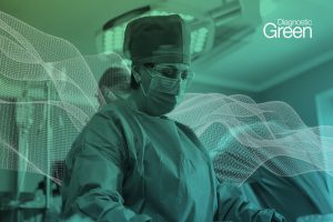
Published in J Laparoendosc Adv Surg Tech, the authors investigated 18 consecutive patients who underwent laparoscopic liver resection for superficial malignant tumors, namely 11 patients with hepatocellular carcinoma (HCC), 5 patients with colorectal liver metastases (CRLM), 1 patient with intrahepatic cholangiocarcinoma (ICC), and 1 patient with thyroid cancer metastasis, using Indocyanine Green (ICG) fluorescence as an adjuvant tool to intraoperative laparoscopic ultrasound (LUS).
Results: An optimal ICG 15-minute clearance retention rate (R15 < 10%) and ICG plasma disappearance rate (<18%/minute) were present in 11 patients (61.1%) and in 14 patients (77.7%), respectively. Liver tumors were 29 in total, including 14 HCCs (48.3%), 13 CRLMs (44.8%), 1 ICC (3.4%), and 1 thyroid cancer metastasis (3.4%). Twenty-nine tumors (100%) were correctly visualized with ICG/fluorescence, as compared with 21 tumors identified with LUS (72.4%). After complete liver mobilization, ICG staining allowed to identify more superficial lesions (early HCC and small CRLM) in posterolateral segments (Segments 6 and 7) as compared with LUS (14 versus 10 lesions). In addition, in segments usually treated laparoscopically (e.g., left lateral segments), ICG was superior to LUS (10 versus 6 lesions) to identify superficial early HCC in patients with macronodular cirrhosis.
Conclusions: ICG visual feedback might substitute the tactile feedback of the hand and might in some cases act as a “booster” of LUS for superficial hepatic lesions.