Robotic-assisted laparoscopic radical prostatectomy represents one of the most common operations in urologic oncology and involves several critical technical steps including pelvic lymph node dissection, cavernous nerve sparing and vesicourethral anastomosis. The quality of performing these steps is linked to functional and oncological outcomes. Indocyanine green [ICG] is a non-radioactive, water-soluble compound which allows for enhanced visualization with near-infrared fluorescence of both anatomical structures and vasculature during complex abdominal operations such as prostatectomy. During the last decade, several investigators have examined the value and role of ICG fluorescence during prostatectomy. In this review, we sought to evaluate the body of evidence for fluorescence-guided robotic prostatectomy as well as assess potential future areas of investigation with this technology.
Laparoscopic duodenum-preserving pancreatic head resection with real-time indocyanine green guidance of different dosage and timing: enhanced safety with visualized biliary duct and its long-term metabolic morbidity
few reports of L-DPPHR are available with alarming bile leakage, and none of them revealed the long-term metabolic outcomes
Continue readingFeatures Found in Indocyanine Green-Based Fluorescence Optical Imaging of Inflammatory Diseases of the Hands
Rheumatologists in Europe and the USA increasingly rely on fluorescence optical imaging (FOI, Xiralite) for the diagnosis of inflammatory diseases. Those include rheumatoid arthritis, psoriatic arthritis, and osteoarthritis, among others. Indocyanine green (ICG)-based FOI allows visualization of impaired microcirculation caused by inflammation in both hands in one examination.
Continue readingThe Use of Indocyanine Green (ICG) and Near-Infrared (NIR) Fluorescence-Guided Imaging in Gastric Cancer Surgery: A Narrative Review
Near-infrared fluorescence imaging with indocyanine green is an emerging technology gaining clinical relevance in the field of oncosurgery. In recent decades, it has also been applied in gastric cancer surgery, spreading among surgeons thanks to the diffusion of minimally invasive approaches and the related development of new optic tools. Its most relevant uses in gastric cancer surgery are sentinel node navigation surgery, lymph node mapping during lymphadenectomy, assessment of vascular anatomy, and assessment of anastomotic perfusion. There is still debate regarding the most effective application, but with relatively no collateral effects and without compromising the operative time, indocyanine green fluorescence imaging carved out a role for itself in gastric resections. This review aims to summarize the current indications and evidence for the use of this tool, including the relevant practical details such as dosages and times of administration.
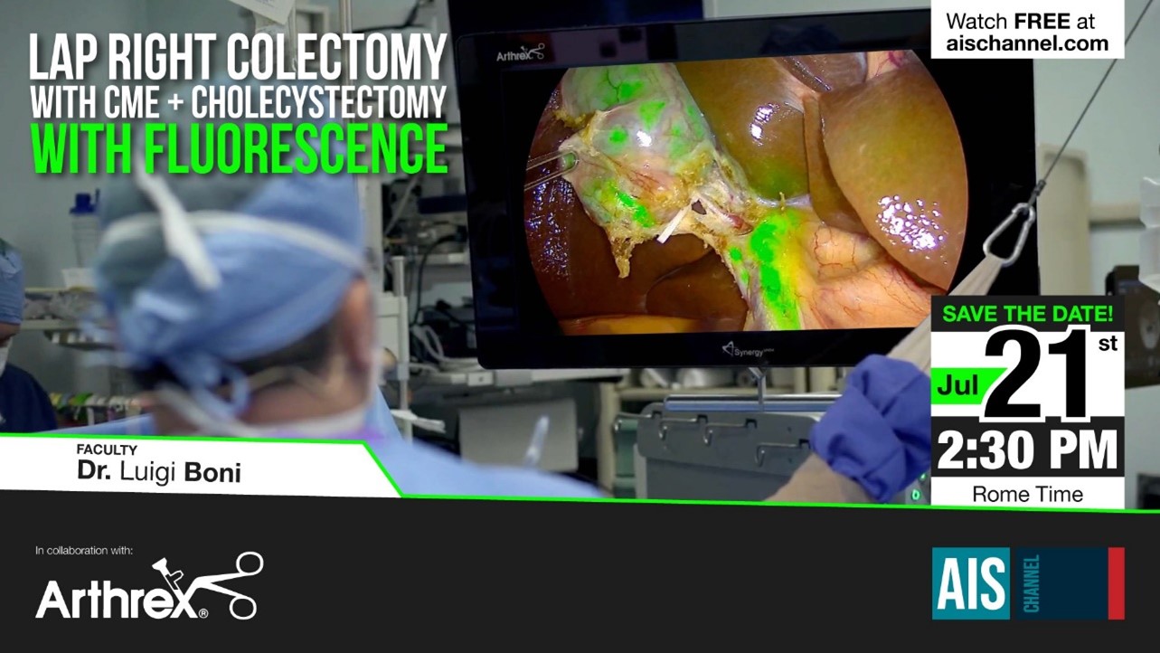
Live Surgery Event on use of ICG in Lap Right Colectomy
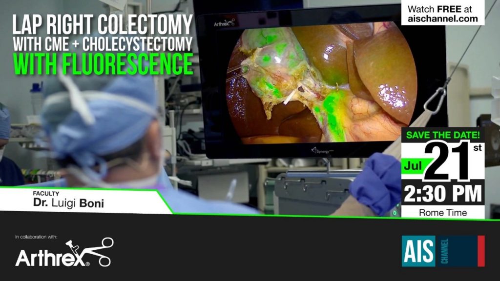
Live Surgery Event on use of ICG in Lap Right Colectomy with Prof Luigi Boni on 21st July 2.30pm Central European Time – Click here to log in/register for free event
July 21 at 02.30pm (Rome Time): Live Surgery: Lap Right Colectomy with CME + Cholecystectomy with Fluorescence
Diagnostic Green are delighted to support this free masterclass on The Role of ICG in Bariatric and Emergency General Surgery and its Role in Education and Training.

Diagnostic Green are delighted to support this free masterclass on The Role of ICG in Bariatric and Emergency General Surgery and its Role in Education and Training.
On 19th July at 7pm BST (8pm CET; 2pm US Eastern Standard Time) we will hear from Prof Boni from Italy, Prof Carus from Germany, Consultant surgeons Mr Awan and Bhatti from UK and Jo Cook from Kimal – Diagnostic Green’s rep in UK. A great and informative webinar promised – don’t miss out.
To register for this unique free event covering Emergency surgery use for ICG, click the following link.
Choroidal changes in posterior microphthalmos
To describe the choroidal variations in posterior microphthalmos (PM). Four eyes of two patients diagnosed as PM based on the characteristic clinical features were included. Multimodal retinal imaging with clinical fundus documentation using ultrawide field fundus camera, optical coherence tomography (OCT) and indocyanine green angiography (ICGA) was done for these cases.
Results: Multimodal imaging of these cases confirmed the variations in the choroid in PM cases. In both cases, on OCT, the retina and choroid were thick. retinal papillomacular fold (RPMF) was noted in all four eyes. On ICGA, the dye transit time from the arm to choroid and retina were within normal limits. Choroidal vasculature in the far retinal periphery was reduced and was noted as hypocyanescent areas anterior to the equator while the density of choroidal vessels was significantly more posterior to the equator. Vortex veins were not visualised in both cases.
Conclusion: Choroidal structure and vessels undergo alterations in PM. Further validation of these findings is required in a larger cohort of PM cases.
Laparoscopic partial nephrectomy for the horseshoe kidney with indocyanine green fluorescence guidance under the modified supine position
Owing to the complexity of their blood supply, renal tumors in horseshoe kidneys are sometimes technically challenging to resect through laparoscopic procedures.
Case presentation: A 75-year-old man presented with a 3-cm lower-pole mass in the right moiety of the horseshoe kidney. Indocyanine green administration allowed for the identification of the tumor’s feeding artery, which was selectively clamped to perform laparoscopic partial nephrectomy. During the procedure, the patient was positioned in the modified supine position (30° semi-lateral position), which enabled us to approach the branch of the left renal artery. Postoperative pathologic examination of the resected mass confirmed the diagnosis of pT1a clear cell renal cell carcinoma with negative surgical margins.
Conclusion: Our novel laparoscopic approach with indocyanine green fluorescence in the modified supine position facilitates the identification of and access to the tumor’s feeding artery. This technique is advantageous for laparoscopic partial nephrectomy in patients with horseshoe kidney.
The Use of near Infra-Red Radiation Imaging after Injection of Indocyanine Green (NIR-ICG) during Laparoscopic Treatment of Benign Gynecologic Conditions: Towards Minimalized Surgery. A Systematic Review of Literature
Background and Objectives: To assess the use of near infrared radiation imaging after injection of indocyanine green (NIR-ICG) during laparoscopic treatment of benign gynecologic conditions.
Materials and Methods: A systematic review of the literature was performed searching 7 electronic databases from their inception to March 2022 for all studies which assessed the use of NIR-ICG during laparoscopic treatment of benign gynecological conditions. Results: 16 studies (1 randomized within subject clinical trial and 15 observational studies) with 416 women were included. Thirteen studies assessed patients with endometriosis, and 3 studies assessed non-endometriosis patients. In endometriosis patients, NIR-ICG use appeared to be a safe tool for improving the visualization of endometriotic lesions and ureters, the surgical decision-making process with the assessment of ureteral perfusion after conservative surgery and the intraoperative assessment of bowel perfusion during recto-sigmoid endometriosis nodule surgery. In non-endometriosis patients, NIR-ICG use appeared to be a safe tool for evaluating vascular perfusion of the vaginal cuff during total laparoscopic hysterectomy (TLH) and robotic-assisted total laparoscopic hysterectomy (RATLH), and intraoperative assessment of ovarian perfusion in adnexal torsion.
Conclusions: NIR-ICG appeared to be a useful tool for enhancing laparoscopic treatment of some benign gynecologic conditions and for moving from minimally invasive surgery to minimalized surgery. In particular, it might improve treatment of endometriosis (with particular regard to deep infiltrating endometriosis), benign diseases requiring TLH and RATLH and adnexal torsion. However, although preliminary findings appear promising, further investigation with well-designed larger studies is needed.
The Role of ICG in Robot-Assisted Liver Resections
Introduction: Robotic-assisted liver surgery (RALS) with its known limitations is gaining more importance. The fluorescent dye, indocyanine green (ICG), is a way to overcome some of these limitations. It accumulates in or around hepatic masses. The integrated near-infrared cameras help to visualize this accumulation. We aimed to compare the influence of ICG staining on the surgical and oncological outcomes in patients undergoing RALS.
Material and methods: Patients who underwent RALS between 2014 and 2021 at the Department of General Surgery at the University Hospital Schleswig-Holstein, Campus Kiel, were included. In 2019, ICG-supported RALS was introduced.
Results: Fifty-four patients were included, with twenty-eight patients (50.9%) receiving preoperative ICG. Hepatocellular carcinoma (32.1%) was the main entity resected, followed by the metastasis of colorectal cancers (17%) and focal nodular hyperplasia (15.1%). ICG staining worked for different tumor entities, but diffuse staining was noted in patients with liver cirrhosis. However, ICG-supported RALS lasted shorter (142.7 ± 61.8 min vs. 246.4 ± 98.6 min, p < 0.001), tumors resected in the ICG cohort were significantly smaller (27.1 ± 25.0 mm vs. 47.6 ± 35.2 mm, p = 0.021) and more R0 resections were achieved by ICG-supported RALS (96.3% vs. 80.8%, p = 0.075).
Conclusions: ICG-supported RALS achieve surgically and oncologically safe results, while overcoming the limitations of RALS.

