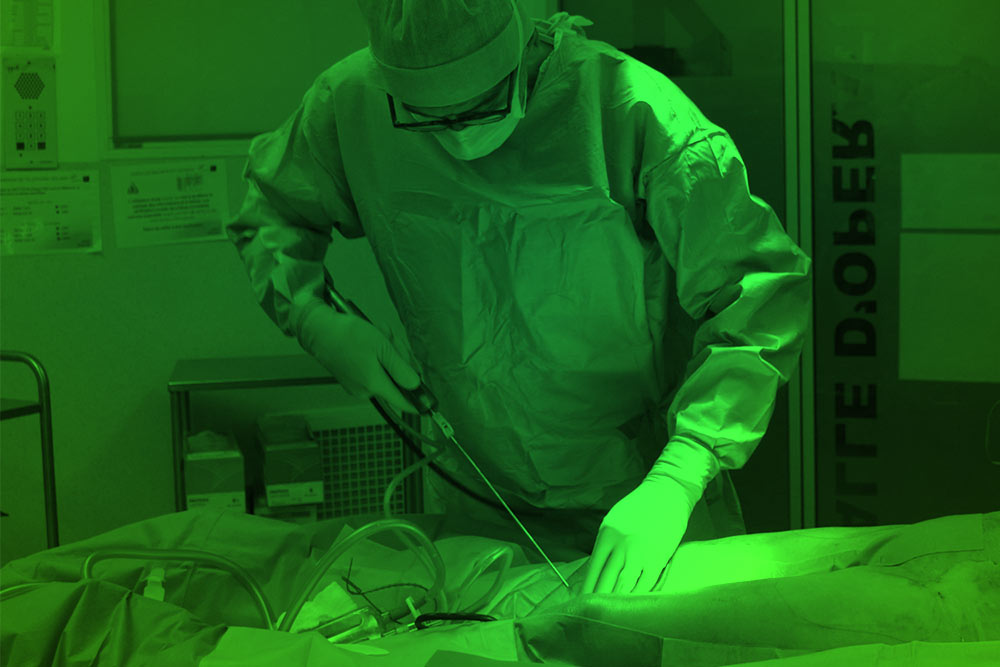
Intestinal hypoperfusion in patients with Crohn’s disease revealed by intraoperative indocyanine green fluorescence imaging

This open access paper notes that

  • Low blood supply increases leakage and surgical recurrence in Crohn’s disease,
  • Indocyanine green angiography is useful for evaluating the intestinal blood supply.
  • Blood supply at anastomotic sites was lower in Crohn’s disease than colon cancer.

Conclusion: Intraoperative indocyanine green fluorescence imaging is safe and reproducible for assessing intestinal perfusion prior to anastomosis in patients with colon cancer and Crohn’s disease.

https://www.sciencedirect.com/science/article/pii/S2049080121003526

Indocyanine green fluorescence angiography versus standard intraoperative methods for prevention of anastomotic leak in colorectal surgery: meta-analysis

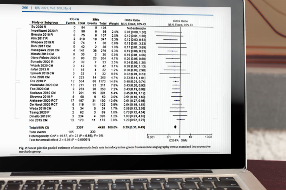
Recent paper published in British Journal of Surgery which undertook an assessment of anastomotic blood perfusion with intraoperative indocyanine green fluorescence angiography (ICG-FA) to evaluate if it may be effective in preventing anastomotic leak compared with standard intraoperative methods in colorectal surgery.

MEDLINE, PubMed, Cochrane Central Register of Controlled Trials and the Cochrane Library were searched for RCTs and observational studies on intraoperative ICG-FA to May 2020.

Results: Twenty-five comparative studies included a total of 7735 patients. The use of intraoperative ICG fluorescence angiography was linked with a significant reduction in all grades anastomotic leak (OR 0.39 (95 per cent c.i. 0.31 to 0.49), P < 0.001; number needed to treat for an additional beneficial outcome (NNTB) 23) and length of hospital stay (MD -0.72 (95 per cent c.i. -1.22 to -0.21) days, P = 0.006). A significantly lower incidence of grade A (OR 0.33 (0.18 to 0.60), P < 0.001), grade B (OR 0.58 (0.35 to 0.97), P = 0.04) and grade C (OR 0.59 (0.38 to 0.92), P = 0.02) anastomotic leak was demonstrated in favour of ICG-FA. For low or ultra-low rectal resection, the odds of developing anastomotic leakage was 0.32 (0.23 to 0.45) (P < 0.001; NNTB 14). There were no differences in duration of surgery, and no adverse events related to ICG fluorescent injection.

Conclusion: The use of ICG-FA instead of standard intraoperative methods to assess anastomosis blood perfusion in colorectal surgery leads to a significant reduction in anastomotic leakage and in the need for surgical reintervention for anastomotic leak, especially in patients with low or ultra-low rectal resections.

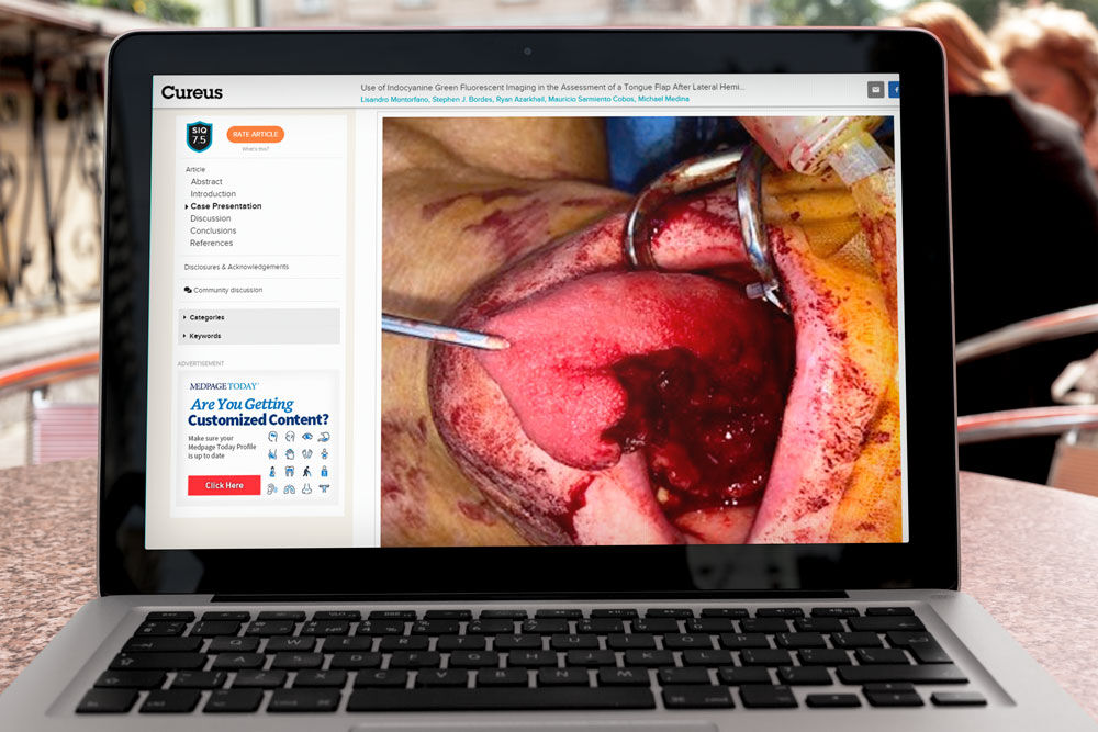
Study published earlier this month “Fluorescence angiography after vascular ligation to make the ileo-anal pouch reach”

Fluorescence angiography after vascular ligation to make the ileo-anal pouch reach

This Dutch study Abstract noted: “The two most essential technical aspects of any gastrointestinal anastomosis are adequate perfusion and sufficient reach. For ileal pouch-anal anastomosis (IPAA), a trade-off exists between these two factors, as lengthening manoeuvers to avoid tension may require vascular ligation. In this technical note, we describe two cases in which we used indocyanine green (ICG) fluorescence angiography (FA) to assess perfusion of the pouch after vascular ligation to acquire sufficient reach. In both cases, ICG FA allowed us to distinguish better between an arterial inflow problem and venous congestion than white light assessment. Both pouches remained viable and no anastomotic leakage occurred. Our results indicate that ICG FA is of great value after vascular ligation to obtain reach during IPAA.”

https://pubmed.ncbi.nlm.nih.gov/33993370/

Case report on Laparoscopic liver cyst fenestration with real-time indocyanine green fluorescence-guided surgery

Case report on Laparoscopic liver cyst fenestration with real-time indocyanine green fluorescence-guided surgery

Abstract: Laparoscopic fenestration (LF) has recently been considered a standard procedure for nonparasitic symptomatic liver cysts. Here, we report a case of LF that was safely performed using real-time indocyanine green (ICG) fluorescence-guided surgery. A 74-year-old woman presented with right upper abdominal pain and poor dietary intake. The patient was diagnosed with symptomatic liver cysts and underwent LF. One hour before surgery, ICG (2.5 mg) was intravenously administered to the patient. ICG fluorescence imaging clearly showed the biliary ducts and distinguished the cysts from the liver parenchyma. We could resect only the cyst walls as wide as possible under the guidance of both white light and fluorescence imaging. There were no signs of postoperative symptom recurrence. Detection of ICG fluorescence in the liver parenchyma is as important as ICG cholangiography for fenestration. Laparoscopic liver cyst fenestration with real-time ICG fluorescence-guided surgery is safe and can be used as a standard procedure.

https://academic.oup.com/jscr/article/2021/5/rjab196/6276408

Recent paper published in Surgery Today “Indocyanine green fluorescence imaging for evaluating blood flow in the reconstructed conduit after esophageal cancer surgery”

Indocyanine-green-fluorescence-imaging-for-evaluating-blood-flow

Journal Abstract: We investigated the effectiveness of indocyanine green (ICG) fluorescence blood flow imaging of the gastric conduit to evaluate anastomotic leakage after esophagectomy. We identified 19 articles using the PRISMA standard for systematic reviews. The more recent studies reported attempts at objective quantification of ICG fluorescence imaging, rather than qualitative assessment. Anastomotic leakage after esophagectomy occurred in 0-33% of the patients who underwent ICG fluorescence imaging. According to the six studies that compared the incidence of anastomotic leakage in the ICG group and the control group, it ranged from 0 to 18.3% in the ICG group and from 0 to 25.2% in the control group, respectively. Overall, the incidence of anastomotic leakage in the ICG group (8.4%) was lower than that in the control group (18.5%). Although the incidence of anastomotic leakage was as high as 43.1% in patients who did not undergo any intraoperative intervention for poor blood flow, it was only 24% in patients who underwent intraoperative intervention. This systematic review revealed that ICG fluorescence imaging may be a crucial adjunctive tool for reducing anastomotic leakage after esophagectomy, suggesting that it should be performed during esophageal reconstruction.

https://pubmed.ncbi.nlm.nih.gov/33977382/

Warram receives NIH R37 MERIT Award

Warram-receives-NIH-R37-MERIT-Award

Jason Warram, Ph.D., associate professor with the University of Alabama at Birmingham Department of Otolaryngology, has received a seven-year NIH R37 MERIT Award from the National Cancer Institute for his research project, “Reduction of Tumor-Positive Margins in Oncologic Surgery.”

In head and neck cancer, tumor-positive margins occur in up to 30 percent of cases, directly correlating with local-regional relapse and poor survival. The high rate of positive margins has not changed in 20 years. Given that the majority of patients with head and neck cancer receive surgery as part of their definitive management, an incremental gain would dramatically impact survival outcomes. By utilizing targeted fluorescent agents for surgery, known as fluorescence-guided surgery, this approach will illuminate head and neck tumor-positive margins during surgery to reduce the rates of recurrence and improve overall surgical outcomes.

Warram holds secondary appointments in the Department of Surgery, Radiology and Biomedical Engineering and is an associate scientist in the O’Neal Comprehensive Cancer Center.

https://www.uab.edu/news/people/item/12037-warram-receives-nih-r37-merit-award

The Efficacy of Intraoperative Fluorescent Imaging Using Indocyanine Green for Cholangiography During Cholecystectomy and Hepatectomy

The-Efficacy-of-Intraoperative-Fluorescent-Imaging-Using-Indocyanine-Green-for-Cholangiography-During-Cholecystectomy-and-Hepatectomy

Japanese paper just published where examined the use of Intraoperative indocyanine green (ICG) cholangiography to help avoid bile duct injuries.

Patients and Methods: We examined 25 patients who underwent intraoperative cholangiography using ICG fluorescence.

Results: There were 24 patients who underwent laparoscopic cholecystectomy and 1 patient who underwent hepatectomy. For laparoscopic cholecystectomy, the average operation time was 127 (50– 197) minutes, and estimated blood loss was 43.2 (0– 400) g. The ICG administration route was intravenous injections in 12 cases and intrabiliary injection in 12 cases (GB injection: 3 cases, PTGBD: 8 cases, ENBD:1 case). The postoperative hospital stay was 4.6 (3– 9) days, and no postoperative complications (Clavien–Dindo ≧IIIa) were observed. For hepatectomy, a tumor located near the left Glissonian pedicle was resected using a fluorescence image guide. Biliary structures were fluorescent without injury after resecting the tumor. No adverse events due to ICG administration were observed, and the procedure was able to be performed safely.

Conclusion: ICG fluorescence imaging allows surgeons to visualize the course of the biliary tree in real time during cholecystectomy and hepatectomy. This is considered essential for hepatobiliary surgery to prevent biliary tree injury and ensure safe surgery.

https://www.dovepress.com/the-efficacy-of-intraoperative-fluorescent-imaging-using-indocyanine-g-peer-reviewed-fulltext-article-CEG

Anesthesiologists Provide Elective Surgery Checklist for Patients During COVID-19

Anesthesiologists-Provide-Elective-Surgery-Checklist-for-Patients-During-COVID-19

As the curve of new COVID-19 cases begins to flatten in the United States, the American Society of Anesthesiologists (ASA) is providing guidelines for patients due for surgery.   

“Physicians, hospitals and health systems are eager to resume elective surgeries and patients are looking to have the procedures they planned, before the pandemic put everything on hold,” said ASA President Mary Dale Peterson, MD, MSHCA, FACHE, FASA. “Health systems can ensure these procedures resume safely by following ASA guidance.”                                      

Partnering with other associations, ASA developed a roadmap to help health care organizations safely resume elective surgeries by assessing readiness, prioritization and scheduling. Patients can use the following checklist to ensure their comfort in moving forward with surgery:

  • Cases are decreasing.
  • All patients are tested.
  • The procedure will be performed in a safe environment.
  • Your physicians are there to answer your questions and concerns.

https://www.newswise.com/coronavirus/anesthesiologists-provide-elective-surgery-checklist-for-patients-during-covid-19

Published in General Surgery News, an interesting article “The Post-Bariatric Patient With Complications”

Published-in-General-Surgery-News,-an-interesting-article-“The-Post-Bariatric-Patient-With-Complications”

Tips on Diagnosis and Management For the Nonbariatric Surgeon

“When patients develop a leak, they will have sustained tachycardia, above 120 [beats per minute] regardless of pain medication or antibiotics. But when the patient has a bleed, their heart rate doesn’t just go up; it also becomes very cyclic, from 80 to 120 to 90 to 110,” said Dr. Rosenthal, the chief of minimally invasive and bariatric surgery at Cleveland Clinic in Weston, Fla.

https://www.generalsurgerynews.com/In-the-News/Article/04-21/The-Post-Bariatric-Patient-With-Complications/63142

Richard Wolf is Corporate Sponsor of the International Society For Fluorescence Guided Surgery (ISFGS)

Richard-Wolf-is-Corporate-Sponsor-of-the-International-Society-For-Fluorescence-Guided-Surgery-(ISFGS)

The medical technology company Richard Wolf GmbH has announced that it has joined up to become a corporate sponsor of the ISFGS (International Society For Fluorescence Guided Surgery).

The ISFGS is dedicated to the global advancement of medical fluorescence imaging through practical application as well as training and the promotion of basic and clinical research. Its goal is to bring together specialists and experts from this field, to produce multimedia training material and thus to strengthen overall awareness and the use of fluorescence imaging. With this partnership, Richard Wolf supports the research and development of this important technology and underlines the claim to promote new medical trends, to promote the exchange of knowledge and thus also to meet the needs and requirements of the users and to offer excellent service.

Richard Wolf GmbH is a medium-sized medical technology company with over 1,500 employees as well as sixteen branches and 130 foreign representatives worldwide. The company develops, produces and sells a variety of products for endoscopy and extracorporeal shock wave treatment in human medicine. Integrated OR systems round off the product portfolio.

https://www.pressebox.de/pressemitteilung/richard-wolf-gmbh/Richard-Wolf-ist-Corporate-Sponsor-der-International-Society-For-Fluorescence-Guided-Surgery-ISFGS/boxid/1056898