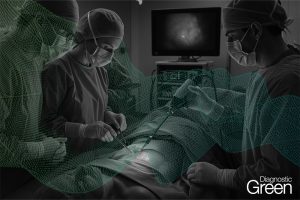
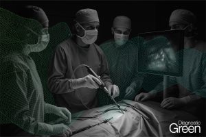
Background: Indocyanine green fluorescence angiography (ICGFA) during colorectal surgery associates with reduced post-operative anastomotic complication rates. Because its interpretation is subjective, quantification has been proposed to address inter-user variability. This study reviews the published literature regarding ICGFA quantification during colorectal surgery with a focus on impactful clinical deployment.
Methods: A systematic review was performed of English language publications regarding clinical studies of ICGFA quantification in colorectal surgery in PubMed, Scopus, Web of Science and Cochrane Library on 29th August 2024, updated to 18th November 2024, following PRISMA guidelines. Newcastle Ottawa scale (NOS) was used to assess quality.
Results: A total of 1428 studies were screened with 22 studies (1469 patients) selected. There was significant heterogeneity of ICGFA methodology, quantification methods and parameter selection and only three studies were NOS “high” quality. Extracorporeal application was most common. Four studies (154 patients) conducted real-time ICGFA analyses (others were post hoc) and four utilised artificial intelligence methods. Eleven studies only included patients undergoing left-sided resection (six focusing specifically on rectal resections). Only one study employed the quantification method to guide intra-operative decision-making regarding colonic transection. Twenty-six different perfusion parameters were assessed, with time from injection to visible fluorescence and maximum intensity the most commonly (but not only) correlated parameters regarding anastomotic complication (n = 18). Other grounding correlates were tissue oxygenation (n = 3, two with hyperspectral imagery), metabolites (n = 2) and surgeon interpretation (n = 5).
Conclusion: Quantification of the ICGFA signal for colorectal surgery is feasible but has so far seen limited academic advancement beyond feasibility.