Use of Verdye (ICG) in
Ophthalmology
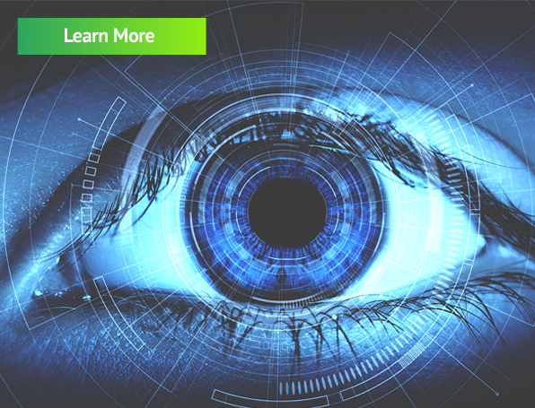
Indocyanine Green Angiography (ICGA) is the gold standard in diagnosing a number of serious eye conditions and is a
key diagnostic tool used by ophthalmology specialists worldwide.
ICGA is particularly useful in the differential diagnosis of Polypoidal Choroidal Vasculopathy (PCV),
Central Serous Chorioretinopathy (CSCR), and Retinal Angiomatous Proliferation (RAP), which can be misdiagnosed
as nAMD (Neovascular Age-related Macular Degeneration)1.
Key Features
Verdye (Indocyanine Green (ICG)) is a tricarbocyanine dye with both hydrophilic and lipophilic properties.
The retention of ICG in the fenestrated choroidal circulation, combined with its low permeability,
makes ICG angiography ideal for viewing the choroidal blood vessels.
Once injected, Verdye binds to plasma proteins and quickly circulates to the choroid layer, delineating the choroidal
veins within 15-20 secs.
Verdye is cleared exclusively through the liver and then excreted through the bile. It does not undergo metabolism. Verdye has an excellent safety profile and adverse reactions occur very rarely (<1/10,000).
Using ICGA at initial presentation helps identify disorders of the choroidal circulation, allowing differential treatment approaches that may improve outcomes and safety
for patients.

- Investigation of complex posterior uveitis and white dot syndromes
- Assessment of patients with “wet” AMD where the presence of polypoidal choroidal vasculopathy (PCV) is in question
- The assessment of choroidal hyperpermeability in patients with central serous chorioretinopathy
For more details go to our FAQ page

ICGA vs OCTA
Prescribing Information
Name of the Medicinal Product
Verdye 5 mg/ml Injection 25 mg / 50 mg, Powder for Solution
Pharmaceutical Form
Powder for Solution for Injection Dark-green powder
Therapeutic indications
This medicinal product is for diagnostic use only.
Method of administration
Before administration the powder must be reconstituted with water for injection.
Shelf Life
5 years. After reconstitution, the solution should be used immediately, protected from light.
Diagnostic Indications
Cardiac, circulatory and micro-circulatory diagnostics
- Measurement of cardiac output and stroke volume
- Measurement of circulating blood volumes
- Measurement of cerebral perfusion
Liver function diagnostics
- Measurement of liver blood flow
- Measurement of excretory function of the liver
Ophthalmic angiography diagnostics
- Measurement of perfusion of the choroid
- Posology and method of administration
Download our Ophthalmology Brochure