The use of ICG in
Animal Health
Verdye for Animal Use 1
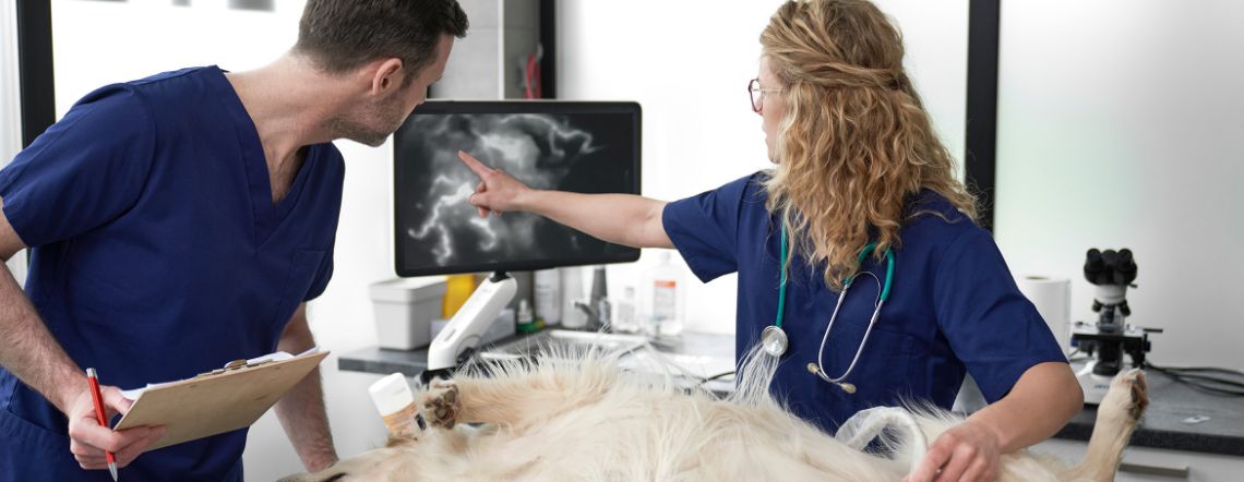
Fluorescence guided surgery in animal health is set to revolutionise the field by providing a non-invasive, real-time and highly specific way to visualise tissues in living animals. It has a wide range of applications in both research and clinical settings, contributing to advancements in visualising anatomy and improving surgical outcomes.2

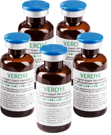
Verdye (Indocyanine Green, ICG) key features and indications for use:
Verdye Injection
Binds to plasma
proteins & quickly
circulates
Fluorescence can
be imaged in
minutes
Cleared by the liver
& eliminated in
the bile
Approved indications (for human use)

Perfusion Assessment
Measurement of cardiac, circulatory and micro-circulatory diagnostics including perfusion

Liver Function Diagnostics
Measurement of excretory function of the liver

Ophthalmic Angiography Diagnostics
Measurement of perfusion of the choroid
Reconstitution
Verdye powder should be reconstituted immediately prior to use by adding 5ml sterile water for injection to the vial containing 25mg of active substance, which is a concentration of 5mg/ml (0.5% w/v). Visually inspect the reconstituted solution. Only use clear solutions free from visible particles. Verdye is a sterile product, intended for single use only.
Administration and dosage
- Diagnostic procedures with Verdye should be performed under the supervision of a veterinary surgeon.
- Verdye is intended for intravenous injection via an injection needle, a central or peripheral catheter or cardiac catheter.
- Verdye binds to plasma proteins and quickly circulates, enabling image capture within minutes.
- To view fluorescence, a Near Infra-Red (NIR) camera is required.


Fluorescence imaging in animal surgery represents a pivotal methodological approach leveraging the utilisation of the fluorescent agent Verdye (ICG) alongside dedicated near-infrared imaging equipment. This advanced modality enables the precise visualisation of targeted tissues and anatomical structures within live animal models. By harnessing near-infrared technology, this surgical approach provides real-time, high-fidelity imagery, during surgery.
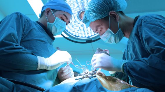
Minimally invasive surgery in animal health has started to expand quickly and has become the preferred technique in many surgical procedures, thanks to its reduced invasiveness and fast postoperative recovery.2
Over the past decade, many clinical applications using Verdye/ICG have been adopted in veterinary medicine. Some of the more commonly performed procedures include liver mass resection, thoracic duct ligation2, sentinel lymph node resection3,6 and other various gastrointestinal surgeries including colon resections.
In human use, the approved indications for Verdye are:

Liver function diagnostics

Ophthalmic Angiography diagnostics

Breast sentinel lymph node biopsy (currently approved in Spain only)
As in human medicine, fluorescence imaging in animal surgery is a valuable technique that involves the use of a fluorescent dye such as Verdye/ICG and specialised camera systems also known as near infrared equipment to visualise specific tissues, structures, or metastases within living animals. This technology provides real-time, high-resolution images that help surgeons and researchers gain insights into the anatomy, physiology and pathology of animals.
Here are some key aspects of fluorescence imaging in animal surgery:

- Intraoperative Guidance: Fluorescence imaging is especially useful in guiding surgeons during procedures. By adopting fluorescence guidance surgery, it highlights important structures or pathological regions resulting in surgeons being able to make more informed decisions in real-time, helping to improve the precision and safety of the surgery.
- Tumour Imaging/Identification: In cancer surgery, fluorescence imaging can be used to visualise tumour margins, identify small metastases and assess the extent of tumour removal. This technique helps surgeons remove cancerous tissue while preserving healthy tissue.4,5
- Blood Flow Visualisation: Verdye/ICG can be injected into the bloodstream to track blood flow patterns in animals. This is particularly useful for monitoring vascular health and assessing the perfusion of tissues during surgery.7
- Real-time Monitoring: Fluorescence imaging provides real-time feedback, allowing surgeons to assess tissue viability, detect abnormalities and make necessary surgery adjustments during surgery.
- Minimally Invasive Procedures: In minimally invasive surgery, such as laparoscopy, fluorescence imaging can help guide the surgeon, thereby improving accuracy and reducing the risk of damage to surrounding tissues.

For further information, download our Use of ICG in Animal Health brochure
References
1. Articles 112, 113 and 114 of European Regulation 2019/6 (the so-called ‘cascade’ provisions), when a suitable veterinary medicinal product is not available in this country, veterinary practitioners are entitled to use certain human medicines under particular circumstances. https://eur-lex.europa.eu/legal-content/EN/TXT/PDF/?uri=CELEX:32019R0006
2. https://vetminimallyinvasivesurgery.edrapublishing.com/
3. Determining agreement between preoperative computed tomography lymphography and indocyanine green near infrared fluorescence intraoperative imaging for sentinel lymph node mapping in dogs with oral tumours. Volume 19Issue 4Veterinary and Comparative Oncology pages: 770-770 First Published online: July 12, 2021
4. Sakurai N, Ishigaki K, Terai K, Heishima T, Okada K, Yoshida O, Kagawa Y, Asano K. Clinical impact of near-infrared fluorescence imaging with indocyanine green on surgical treatment for hepatic masses in dogs. BMC Vet Res. 2022 Oct 19;18(1):374. doi: 10.1186/s12917-022-03467-2. PMID: 36261863; PMCID: PMC9580212.
5. Nakaseko Y, Ishizawa T, Saiura A. Fluorescence-guided surgery for liver tumors. J Surg Oncol. 2018 Aug;118(2):324-331. doi: 10.1002/jso.25128. Epub 2018 Aug 11. PMID: 30098296.
6. Muhanna N, Chan HHL, Douglas CM, Daly MJ, Jaidka A, Eu D, Bernstein J, Townson JL, Irish JC. Sentinel lymph node mapping using ICG fluorescence and cone beam CT – a feasibility study in a rabbit model of oral cancer. BMC Med Imaging. 2020 Sep 14;20(1):106. doi: 10.1186/s12880-020-00507-x. PMID: 32928138; PMCID: PMC7491106
7. Adam S. F. Quinlan BScH, Shannon H. Wainberg DVM, Erin Phillips DVM, BSc, Michelle L. Oblak DVM, DVSc, DACVS, ACVS. The use of near infrared fluorescence imaging with indocyanine green for vascular visualization in caudal auricular flaps in two cats. First published: 25 January 2021
