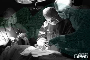
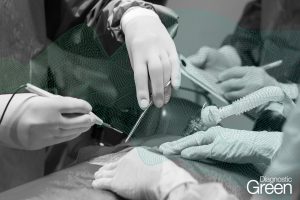
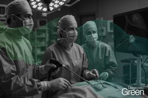
The integration of indocyanine green (ICG) fluorescence imaging into robotic liver segmentectomies has improved intraoperative visualization for parenchymal-sparing hepatic resections. This systematic review and meta-analysis of 15 prospective and retrospective studies (n = 612 patients) found that ICG navigation was associated with higher rates of R0 resection, with a pooled odds ratio of 2.34 (95% CI 1.56-3.51, I2 = 32%) compared with conventional techniques.
Quantitative analysis showed reductions in intraoperative blood loss (mean difference – 85 mL, 95% CI – 120 to – 50 mL) and bile leak incidence (3.1% vs 6.1%, p = 0.03), with no statistically significant difference in operative times (mean difference – 12 min, p = 0.12). Subgroup analyses indicated greater benefits for colorectal liver metastases (OR 3.12, 95% CI 1.89-5.15) and posterosuperior segment resections (margin improvement + 2.3 mm, p = 0.03). Efficacy was lower in cirrhotic livers (82.4% vs 97.1% success rate, p = 0.002), suggesting a need for tailored protocols in this group. Reported protocols varied by surgical aim: systemic 24-h preoperative injection was primarily used for tumor localization and margin visualization, while intraoperative portal vein injection was applied for segmental demarcation using positive or negative staining.
These findings support ICG fluorescence as a useful adjunct in robotic liver segmentectomies, while underscoring the need to standardize dosing regimens and evaluate long-term survival outcomes.