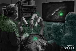
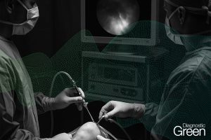
Reported case study where patient initially underwent the endoscopic retrograde cholangiopancreatography (ERCP) and endoscopic sphincterotomies (EST) with stents placement on the admission day, followed by an early laparoscopic cholecystectomy on the second day. The indocyanine green (ICG) fluoresced imaging system was applicated in the surgery. During the surgery, the difficult identification of the gallbladder anatomy was encountered. Due to severe inflammation and extensive adhesions the standard critical view of safety (CVS) could not be achieved.
According to the current clinical research evidence, early laparoscopic cholecystectomy within the same hospital admission for patients with mild to moderate acute biliary pancreatitis could reduce the risk of recurrent biliary events without increasing perioperative morbidity.
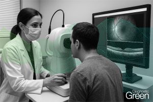
The ICG fluoresced imaging is now general application in surgery particularly in hepatobiliary surgery. In this case, although the Calot’s triangle could not be seen, the border between liver and gallbladder could be well identified with the ICG-fluorescence imaging.
https://www.sciencedirect.com/science/article/pii/S1015958423015567?via%3Dihub