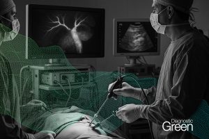
Near infrared (NIR) analysis of tissue perfusion via indocyanine green fluorescence assessment is performed clinically during surgery for a range of indications. Its usefulness can potentially be further enhanced through the application of interpretable artificial intelligence (AI) methods to improve dynamic interpretation accuracy in these and also open new applications. While its main use currently is for perfusion assessment as a tissue health check prior to performing an anastomosis, there is increasing interest in using fluorophores for cancer detection during surgical interventions with most research being based on the paradigm of static imaging for fluorophore uptake hours after preoperative dosing.
Although some image boosting and relative estimation of fluorescence signals is already inbuilt into commercial NIR systems, fuller implementation of AI methods can enable actionable predictions especially when applied during the dynamic, early inflow-outflow phase that occurs seconds to minutes after ICG (or indeed other fluorophore) administration.
Already research has shown that such methods can accurately differentiate cancer from benign tissue in the operating theatre in real time in principle based on their differential signalling and could be useful for tissue perfusion classification more generally. This can be achieved through the generation of fluorescence intensity curves from an intra-operative NIR video stream. These curves are processed to adjust for image disturbances and curve features known to be influential in tissue characterisation are extracted. Existing machine learning based classifiers can then use these features to classify the tissue in question according to prior training sets.
The use of this interpretable methodology enables accurate classification algorithms to be built with modest training sets in comparison to those required for deep learning modelling in addition to achieving compliance with medical device regulations. Integration of the multiple algorithms required to achieve this classification into a desktop application or medical device could make the use of this method accessible and useful to (as well as useable by) surgeons without prior training in computer technology. This document details some technical and functional design considerations underlying such a novel recommender system to advance the foundational concept and methodology as software as medical device for in situ cancer characterisation with relevance more broadly also to other tissue perfusion applications.