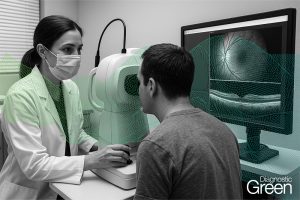
Liver resection is the best treatment for hepatocellular carcinoma (HCC) when resectable. Unfortunately, many patients with HCC cannot undergo liver resection. Percutaneous thermoablation represents a valid alternative for inoperable neoplasms and for small HCCs, but it is not always possible to accomplish it. In cases where the percutaneous approach is not feasible (not a visible lesion or in hazardous locations), laparoscopic thermoablation may be indicated. HCC diagnosis is commonly obtained from imaging modalities, such as CT and MRI, However, the interpretation of radiological images, which have a two-dimensional appearance, during the surgical procedure and in particular during laparoscopy, can be very difficult in many cases for the surgeon who has to treat the tumor in a three-dimensional environment.
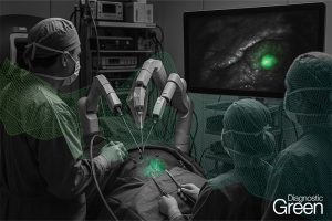
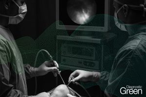
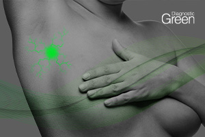
In recent years, more technologies have helped surgeons to improve the results after ablative treatments. The three-dimensional reconstruction of the radiological images has allowed the surgeon to assess the exact position of the tumor both before the surgery (virtual reality) and during the surgery with immersive techniques (augmented reality). Furthermore, indocyanine green (ICG) fluorescence imaging seems to be a valid tool to enhance the precision of laparoscopic thermoablation. Finally, the association with laparoscopic ultrasound with contrast media could improve the localization and characteristics of tumor lesions. This article describes the use of hepatic three-dimensional modeling, ICG fluorescence imaging and laparoscopic ultrasound examination, convenient for improving the preoperative surgical preparation for personalized laparoscopic approach.