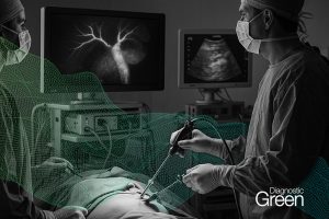
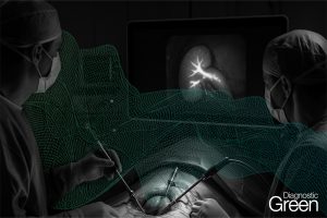
Perforator imaging is a prerequisite in preoperative planning of the peroneal perforator flap and the fibula skin island. Although reports indicate that indocyanine green fluorescence imaging assessment method might be advantageous over conventional ultrasound-based techniques (i.e., Doppler and color duplex), in practice, clear evidence is lacking. Thus, a comparative assessment of the utility of indocyanine green angiography and ultrasound-based techniques in the identification of suitable lower leg skin perforators was performed.
Methods: A prospective clinical cohort study with a series of 12 consecutive patients was conducted to assess indocyanine green angiography, Doppler ultrasound and color duplex ultrasound techniques for preoperative perforator detection in the lower leg before free fibula flap harvest. Anatomical dissection served as a reference. Parameters measured were perforator spatial distance to the reference (precision), operative time expenditure, and ease of device usage for assessment/outcomes.
Results: This study included 12 patients, with a total of 27 perforators. Exhibition of technique sensitivity and positive predictive values were as follows: indocyanine green angiography, 93 percent and 100 percent; Doppler ultrasound, 82 percent and 82 percent; and color duplex ultrasound,89 percent and 86 percent, respectively. With regard to the indocyanine green angiography technique, the distance to the actual perforator location was significantly shorter, which aided detection and lesser time expenditure during operation.
Conclusions: The indocyanine green angiography technique proved to have high precision, sensitivity, positive predictive value, and easy-to-use capabilities because of its exceptional spatial and temporal information, compared to the conventional, ultrasound-based techniques. Therefore, indocyanine green angiography is superior for preoperative perforator imaging of the lateral lower leg.