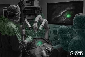
Aim: Indocyanine green video angiography (ICG-VA) effortlessly and immediately allows vascular structures imagination during micro-neurosurgical operations. In this study, we present our experience of 44 consecutive carotid endarterectomy procedures in 42 patients and assess the efficacy and success of ICG-VA in the localization of the plaque sites, extent of the arteriotomy, evaluation of the flow, and presence of thrombus after closure.
Results: Forty-two consecutive patients who underwent a total of 44 CEAs were included. The population consisted of 5 (11.9%) female and 37 (88.1%) male patients, all of whom had at least 60% carotid stenosis, as assessed using North American Symptomatic Carotid Endarterectomy Trial stenosis ratios. The mean stenosis rate was 80.55% (range, 60%-90%), the mean patient age was 69.8 years (range, 44-88 years), and the mean follow-up duration was 40 months (range, 2-106 months). In 31 (70.5%) of 44 procedures, ICG-VA revealed the exact location of the obstructive plaque’s distal end, and it successfully showed the arteriotomy length, identifying the location of the plaque. ICG-VA correctly evaluated the flow in 38 (86.4%) of 44 procedures.
Conclusion: Our reported study is cross-sectional, reflecting our experiment using ICG during CEA. ICG-VA can be used as a simple, practical, real-time microscope-integrated technique that can enhance the safety and effectiveness of CEA.