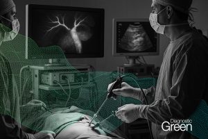
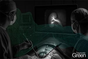
Background: Surgeons can minimize the risk of bile duct injury (BDI) during challenging mini-invasive cholecystectomy through technical standardization by means of a precise anatomical landmark identification (Critical View of Safety) and advanced technology for biliary visualization. Among these systems, the adoption of magnified stereoscopic 3-dimensional view provided by robotic platforms and near infrared fluorescent cholangiography (NIRF-C) is the most promising.
Methods: In this prospective cohort study, we evaluated all consecutive minimally invasive cholecystectomies (laparoscopic and robotic) performed with NIRF-C between May 2022 and January 2023 at General Surgery Unit, Department of Health Sciences, University of Milan, San Paolo Hospital (Milan, Italy). Inclusions criteria were as follows: (1) acute cholecystitis (emergency group), (2) history of chronic cholecystitis or complicated cholelithiasis (deferred urgent group), (3) difficult cases (patients affected by cirrhosis, with scleroatrophic gallbladder or BMI > 35 kg/m2). For each group, the detection rate and visualization order of the main biliary structures were reported (cystic duct, CD; common hepatic duct, CHD; common bile duct, CBD; and CD-CHD junction).
Results: A total of 101 consecutive patients were enrolled, including 83 laparoscopic and 18 robotic cholecystectomies. All patients were stratified into three subgroups: (a) emergency group (n = 33, 32.7%), (b) deferred urgent group (n = 46, 45.5%), (c) difficult group (n = 22, 21.8%). Visualization of at least one biliary structure was possible in 94.1% of cases (95/101). Interestingly, all four main structures were detected in 43.6% of cases (44/101). The CD was the structure identified most frequently, being recognized in 91/101 patients (90.1%), followed by CBD (83.2%), CHD (62.4%), and CD-CHD junction (52.5%). In the subset of patients that underwent emergency surgery for AC, the CD-CHD confluence was identified in only 45.5% of cases. However, early and precise identification of CBD (75.8%) and CD (87.9%) allowed safe isolation, clipping, and transection of the cystic duct. In the deferred urgent group, the CBD and the CD were easily identified as first structure in a high percentage of cases (65.2% and 41.3% respectively), whereas the CD-CHD junction was the third structure to be identified in 67.4% of cases, the highest value among the three subgroups. In the difficult group, NIRF-C did not prove to be a useful tool for biliary visualization. The rates of failure of visualization were elevated: CBD (27.3%), CD (18.2%), CHD (54.5%), and CD-CHD (68.2%).
Conclusions: NIRF-C is a powerful real-time diagnostic tool to detect CBD and CD during minimally invasive cholecystectomy, especially when inflammation due to acute or chronic cholecystitis subverted the anatomy of the hepatoduodenal ligament.